Tumours of the CNS are the second cause of death among children in developed countries after leukaemia, with solid tumours being more common at this age.
In Spain, according to the National Tumour Register, every year 1,500 new cases of infantile cancer, are recorded, of which 20% correspond to tumours of the Central Nervous System.
The annual incidence of CNS tumours in children is 5/100,000, which means 100 new cases a year in Spain.
Infratentorial tumours (55%) predominate in children, that is ones located below the tentorium cerebelli, a membrane that separates the skull into two compartments, far more than supratentorial tumours (45%), except in nursing infants where the supratentorial location predominates.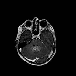
Aetiology:
The causes of brain tumours in children can be attributed to different factors:
- Disorders in CNS formation in many congenital tumours.
- Neurocutaneous syndromes. Chromosomal abnormalities that increase susceptibility to the appearance of tumours throughout life.
- Other genetic disorders and family susceptibility.
- Exogenous factors: radiation, etc.
Clinical signs
The diagnosis of brain tumours in children is often very difficult due to the lack of collaboration and the inability to describe the symptoms due to their age. In older children, collaboration in the clinical record and physical examination mean that the diagnosis can be established much more easily.
In children under two years of age, the detection of macrocephaly, a skull larger than expected for their age, should indicate the suspected presence of hydrocephalus or a cerebral tumour. Macrocephaly is the main indication, although not specific, of a cerebral tumour due to growth of the head as a result of the tumour itself or secondary hydrocephalus. At these early ages the bones of the skull have not yet solidified and it may increase in size to compensate for the increased pressure caused by the tumour or hydrocephalus.
In older children, when the skull is rigid the signs and symptoms of intracranial hypertension are sustained headache, vomiting and papilloedema. The appearance of these symptoms should alert the family and be a reason to consult a physician.
These children may present focal neurological deficits, loss of strength in an extremity, speech difficulties, etc. depending on the cerebral location of the tumour and epilepsy.
Other less specific symptoms are character changes and delayed psychomotor development and growth.
Diagnosis:
A detailed clinical record and physical examination are the basis for any diagnosis in medicine. This information is complemented by imaging tests performed on the central nervous system. The three most important tests in children are:
- Transfontanelle ultrasonography: a non-invasive test based on ultrasounds that enable the intracranial study of children with the fontanelle still open. It enables rejecting hydrocephalus and other causes of macrocephaly in these children.
- 2. Computed tomography (CT): a fast diagnostic test that provides general information on whether there is or not a tumour, its location, extension and characteristics. It is a readily available rapid test and in many cases may be performed without any need for sedation. It enables a first approximation to the presence of brain lesions. In the event of confirming or suspecting the presence of lesions the study should be completed with more selective tests such as brain magnetic resonance imaging, arteriography, etc.
- 3. Magnetic resonance imaging (MRI): this imaging test provides the same information as the CT scan but with much greater detail and without irradiating the child. The inconvenience is that it is not readily available and it requires a longer period of immobility, this is why sedation is often required.
Treatment
The basic treatment for these lesions is surgery, as it meets the double purpose of diagnosis and treatment.
Surgery enables obtaining tumour tissue samples for analysis, typing and a decision regarding the most suitable treatment.
It also enables resecting or reducing the lesion, thus ensuring greater efficacy of other treatments whenever necessary.
The approach to this pathology must be multidisciplinary. And on many occasions it also involves Oncologists and Radiotherapists.
Hydrocephalus due to the compression of the tumour itself is often the cause of the clinical decompensation and so this condition must also be treated in addition to the tumour.