These lesions are benign and originate in ectodermal epithelial tissue. They are also known as pearl tumour (because of its pearly colour and lack of vascularisation) and/or cholesteatomas.
They represent 1% of all intracranial tumours. The most common location in the central nervous system is the cerebellopontine angle. They also appear on a spinal column level.
Clinical signs
 The signs may be those of intracranial hypertension (headache, nauseas, vomiting, dizziness, etc.) and depending on the location may cause different neurological alterations. It is also possible that they cause repetitive meningitis.
When located in the cerebellopontine angle, they may cause trigeminal neuralgia or affect other cranial pairs (facial mobility, swallowing, etc.)
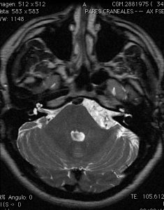
Diagnostic tests
Magnetic resonance imaging is essential for correct assessment of the lesion and to determine its anatomical relationship to surrounding tissues.
Treatment
The treatment of choice is surgical. Correct planning through neuronavigation and experience in neurophysiological monitoring is essential to reduce the risks of surgery (especially when cranial pairs are affected).